


Introduction
Medical photography in maxillofacial surgery is one of PTD’s key services, providing accurate documentation of treatment procedures, functional and aesthetic outcome evaluation, and supporting scientific education and documentation. This service is performed using professional equipment, standard protocols, and a trained team, before, during, and after surgery.

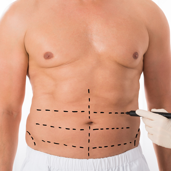
Clinical applications
- Recording the patient’s anatomical position before surgery
- Documenting surgical procedures to review techniques
- Evaluation of treatment results after surgery
- Support for custom prosthetic design and virtual surgical planning
- Providing scientific images for education, research, and publication of articles
Assisting in the precise design of prosthetics and surgical guides
Providing realistic images for training surgeons and students
Review of functional and aesthetic results over time
Benefits
Transparent recording of treatment stages for legal and insurance purposes
Showing the treatment process and increasing the patient's trust in the treatment team
Features
Ordering and design process
Product technical specifications
DSLR cameras with 85mm and 105mm portrait lenses
Ring lights or studio flashes with constant color temperature
High-resolution JPEG and RAW
Frontal, Profile, Oblique, Basal, Smile
Neutral color (gray or blue) with uniform light
Encrypted storage accessible to the treatment team
Clinical Studies and Scientific Support
According to the DTJournal’s expert guide and a study published in Springer, adhering to the principles of medical photography in maxillofacial surgery plays an important role in increasing the accuracy of treatment, scientific documentation and improving education. The images captured by the PTD team comply with international standards and can be used in digital design, scientific articles and medical records.